| Table of Contents |  |
|
Case Report
| ||||||
| Right sided diaphragmatic hernia of Morgagni with associated anatomical abnormalities | ||||||
| Michelle Annabi1, Kristen Farraj1, Melinda Danowitz1, Nikos Solounias2 | ||||||
|
1Medical student, Anatomy, New York Institute of Technology College of Osteopathic Medicine, Old Westbury, NY, USA
2Professor, Anatomy, New York Institute of Technology College of Osteopathic Medicine, Old Westbury, NY, USA | ||||||
| ||||||
|
[HTML Abstract]
[PDF Full Text]
[Print This Article]
[Similar article in Pumed] [Similar article in Google Scholar] |


| How to cite this article |
| Annabi M, Farraj K, Danowitz M, Solounias N. Right sided diaphragmatic hernia of Morgagni with associated anatomical abnormalities. Edorium J Anat Embryo 2017;4:1–5. |
|
ABSTRACT
|
|
At New York Institute of Technology College of Osteopathic Medicine, a rare type of diaphragmatic hernia has been observed within a cadaver in the Anatomy department during routine dissections. Normally, congenital diaphragmatic hernias appear on the left side, namely a Bochdalek hernia. This particular hernia, a Morgagni type, is uniquely found on the anterior right side of the thorax. This 101- year-old female cadaveric specimen had a herniation of the greater omentum, stomach, and portion of the duodenum into the right side of the thorax, with an associated slight mediastinal shift to the left. In addition, this cadaver had a fleshy ‘string’ of tissue connecting the hernia sac to the sigmoid colon, a tortuous internal carotid artery, thickened costal cartilages, and weak fascia.
| |
|
Keywords:
Congenital malformation, Diaphragmatic hernia, Morgagni hernia, Tortuous artery
| |
|
INTRODUCTION
| ||||||
|
The central tendon (composed from the septum transversum), pleuroperitoneal membranes, muscle, and mesentery of the esophagus (which forms the crura of the diaphragm) all come together to form the diaphragm. The development of the diaphragm begins with the septum transversum separating the primitive thoracic and peritoneal cavities [1]. In early embryonic stages, the septum transversum does not completely close off the thoracic cavity. Within the thorax, there are pericardioperitoneal cavities on each side of the foregut where the lungs grow. As development continues, these cavities are closed up by pleuroperitoneal folds and eventually fuse with the mesentery of the esophagus and the septum transversum [2][3] . A hernia occurs when an organ (or organs), is displaced from its original internal location and protrudes through a weakness in the surrounding body cavity. A diaphragmatic hernia, specifically, describes the protrusion of abdominal viscera into the thorax through a weakness in the diaphragm. There are various forms of congenital diaphragmatic hernias that are categorized depending on where and how they travel through the diaphragm. The Bochdalek hernia is postulated to arise when the pleuroperitoneal folds do not fuse with the transverse septum and the dorsal mesentery of the esophagus, therefore creating a posterolateral defect in the diaphragm, allowing for herniation [4]. Morgagni hernias can form when there is a defect in the fusion of the pars sternalis to the costal arches, and therefore are positioned more anteriorly and on the right [5]. The Morgagni hernia is considered to be more rare than the Bochdalek hernia, only presenting in around 2–3% of congenital diaphragmatic hernias [4]. | ||||||
|
CASE REPORT
| ||||||
|
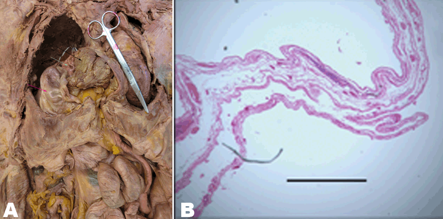
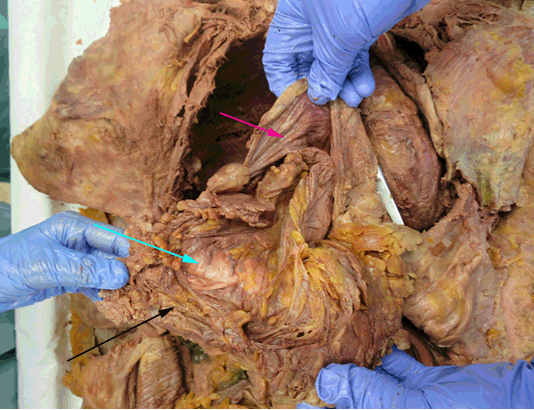
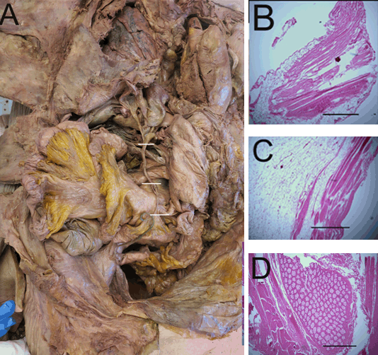
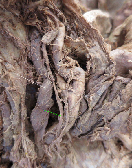
During a routine dissection of the thorax of a 101-year-old female cadaver at New York Institute of Technology College of Osteopathic Medicine, a rare and unusual herniation of abdominal organs through the diaphragm was observed. There was a herniation of greater omentum, stomach, and a portion of the duodenum in a sac above the diaphragm and below the right lung (Figure 1A). The hernial sac was composed of diaphragmatic tissue (Figure 1B). The costal cartilages were raised and thickened. Besides a slight mediastinal shift to the left, the lungs and heart appeared normal. In the abdomen, there was no greater omentum covering the organs because it was displaced in the hernia. When the omentum was removed from the herniated sac, it was noted to be of normal size and without necrosis or atrophy (Figure 2). There was an additional thin, elongated ‘string’ attaching the herniated sac to the sigmoid colon. The ‘string’ was thin, pink, and showed no signs of necrosis (Figure 3). Histological samples of anomalous string were taken and examined. Three samples of the ‘string’ described the internal structure of the string: the most proximal segment- attaching to the hernia, the middle segment, and lastly the distal portion attached to the sigmoid colon. The more proximal part of the ‘string’ resembled features of muscle and fibrous tissue, which is commonly found in the diaphragm (Figure 3B). The middle segment displayed features of muscles, vessels, and adipose omentum (Figure 3C). The distal portion exhibited entrapped intestine and congested blood vessels (Figure 3D). We found that this cadaver had weak fascia. For example, this cadaver had a prolapsed uterus and overall muscle atrophy. It was also observed that this cadaver did not have an observable external urethral sphincter and glans clitoris. In addition, she did not have a left parotid gland and her left internal carotid artery was very tortuous (Figure 4). Other than the herniated abdominal viscera and connection with the sigmoid colon, the abdominal organs appeared grossly normal (Figure 1). | ||||||
|
| ||||||
| ||||||
|
| ||||||
| ||||||
|
DISCUSSION
| ||||||
|
As many as 1 in 2000 babies are born with a congenital diaphragmatic hernia [7], and approximately 85–90% of those cases have a herniation on the left. This condition was likely congenital, and this patient most likely lived asymptomatically. In this specimen, the costal cartilages were raised, presumably due to the lack of space in the thorax. This physical compensation for the decreased thoracic size likely indicates that the herniation must have occurred at a young age. Diaphragmatic herniations have the potential to cause additional health concerns, such as pulmonary hypertension [8][9]. This occurs due to an increase pressure in the thorax from the organs of the abdomen taking up space in the chest. As stated above, in this cadaver, the left internal carotid artery was notably torturous. This observation can be linked to aging, hypertension, and genetic defects [10]. All of these factors are plausible to this case since she was 101-year-old, and medical records indicated that the patient suffered from hypertension. Depending on the level of tortuosity it could have been asymptomatic or cause severe symptoms such as ischemic attacks or stroke. However, the cadaveric brain appeared grossly normal, and without signs of large infarcts. Along with this patient’s age, these symptoms might have led to her death. It was also observed that this cadaver had weak fascia, but this could be attributed to her age. While the weakening of the fascia might have caused the diaphragmatic hernia [11], it is more likely that the defect was congenital and preceded the fascial defects, as acquired diaphragmatic hernias are rare [12]. In addition, she was most likely bed-bound, which was evident from areas of skin breakdown on the lower extremities and buttocks. The anomalous ‘string’ of material attaching the hernial sac to the sigmoid colon likely was a portion of the diaphragm encasing the part of intestine that has traveled with the sac. The ‘string’ may have been a way of transportation for blood and nutrients to the hernia, evidenced by the histologic slides showing the presence of blood vessels. These findings support our observation that the sac originated embryologically in the abdominal region and became entrapped by diaphragm during development. When observing the histology of the sac that was found above the diaphragm, there were no anomalies present and seemed to be muscle similar to the diaphragm encasing all the content. After an extensive review of all the anomalies present in this cadaver and the histological studies it seems that this hernia would be considered to be that of the Morgagni type. The described hernia in our cadaver was limited to the right side of the thorax and was positioned anteriorly, with an associated right sided diaphragmatic defect. | ||||||
|
CONCLUSION
| ||||||
|
A Morgagni type hernia has been identified in this case report. Unlike other diaphragmatic hernias, this hernia is normally located on the right side and is more anterior relative to other hernias. Normally, congenital hernias are associated with other anomalies. In this case, weak fascia, a fleshy ‘string’ spanning the abdomen, and a tortuous carotid artery were noted. Interestingly, the herniated organs and the ‘string’ traveling with it were functional, and the cadaver lived a long life of 101 years. | ||||||
|
REFERENCES
| ||||||
| ||||||
|
SUGGESTED READING
| ||||||
| ||||||

|
[HTML Abstract]
[PDF Full Text]
|
|
Author Contributions
Michelle Annabi – Substantial contributions to conception and design, Drafting the article, Revising it critically for important intellectual content, Final approval of the version to be published Kristen Farraj – Substantial contributions to conception and design, Drafting the article, Revising it critically for important intellectual content,, Final approval of the version to be published Melinda Danowitz – Contributions to conception and design, Drafting the article, Revising it critically for important intellectual content,, Final approval of the version to be published Nikos Solounias – Contributions to conception and design, Drafting the article, Revising it critically for important intellectual content, Final approval of the version to be published |
|
Guarantor of submission
The corresponding author is the guarantor of submission. |
|
Source of support
None |
|
Conflict of interest
Authors declare no conflict of interest. |
|
Copyright
© 2017 Michelle Annabi et al. This article is distributed under the terms of Creative Commons Attribution License which permits unrestricted use, distribution and reproduction in any medium provided the original author(s) and original publisher are properly credited. Please see the copyright policy on the journal website for more information. |
|
|