| Table of Contents | |
|
Case Report
| ||||||
| Atlanto-occipital fusion: A rare anomaly of the craniocervical junction | ||||||
| Dipti A. Nimje1, Harish A. Wankhede2 | ||||||
|
1MD, Anatomy, Medical Officer, Government Medical College, Nagpur, Maharashtra, India.
2MD, Anatomy, Assistant Professor, Department of Anatomy, Government Medical College, Miraj, Maharashtra, India. | ||||||
| ||||||
|
[HTML Abstract]
[PDF Full Text]
[Print This Article]
[Similar article in Pumed] [Similar article in Google Scholar] |
| How to cite this article |
| Nimje DA, Wankhede HA. Atlanto-occipital fusion: A rare anomaly of the craniocervical junction. Edorium J Anat Embryo 2014;1:1–4. |
|
Abstract
|
|
Atlanto-occipital joint is important joint present at the craniocervical junction. This joint comes in relation to the important structures in the region like vertebral arteries, spinal arteries, medulla oblongata and spinal cord. Atlanto-occipital fusion is a rare anomaly of the craniocervical junction which may be congenital or acquired. Clinical symptoms due to this anomaly vary from simple pain to sudden death. So aim of presenting this paper is to aware the clinicians, surgeons, radiologists of such rare anomaly which can land up in severe complications.
| |
|
Keywords:
Atlanto-occipital fusion, Atlas vertebra, Occipitalization, Assimilation, Synostosis
| |
|
Introduction
| ||||||
|
The atlanto-occipital joint is a paired ellipsoid type of synovial joint. Each joint consists of two reciprocally curved articular surfaces. Stability of the joint is maintained by the fibrous capsule, the reciprocally curved articular facets, atlanto-occipital membrane, muscles on the posterior aspect of the neck and other ligaments connecting the bones. Posteriorly, the joints are closely related to the vertebral arteries and dorsal primary ramus of the first cervical nerve and they are the 'at risk' structures present in this region [1]. Other structures such as medulla oblongata, spinal cord and spinal arteries are also vulnerable to damage. The fusion of atlas vertebra with occipital bone is known by various names like occipitalization [2], assimilation of the atlas [3], occipitocervical synostosis [4]. As quoted by Khamanarong et al. the incidence of the atlanto-occipital fusion varies from 0.14–3.63% [2]. Atlanto-occipital fusion is a rare anomaly which may be congenital or acquired [5]. The aim of this paper is to present a rare anomaly, atlanto-occipital fusion and discuss the similar cases found by other researchers. | ||||||
|
Case Report
| ||||||
|
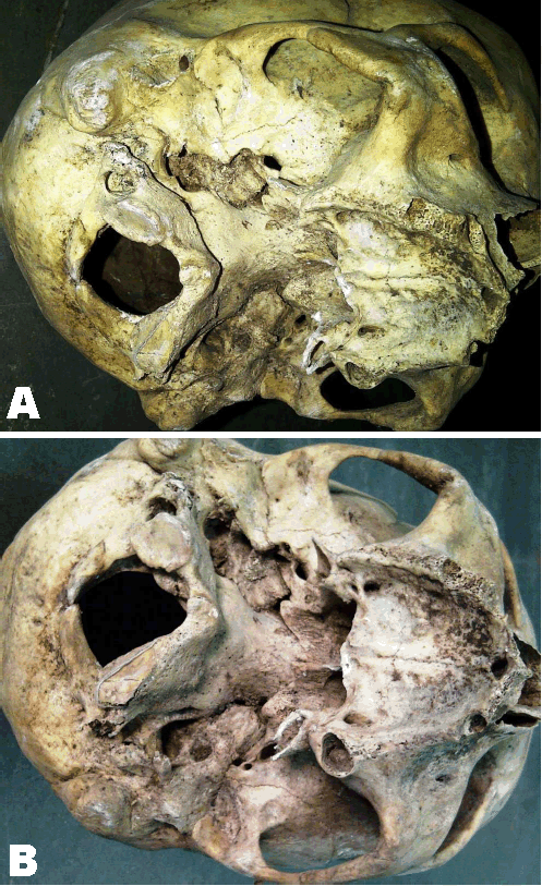
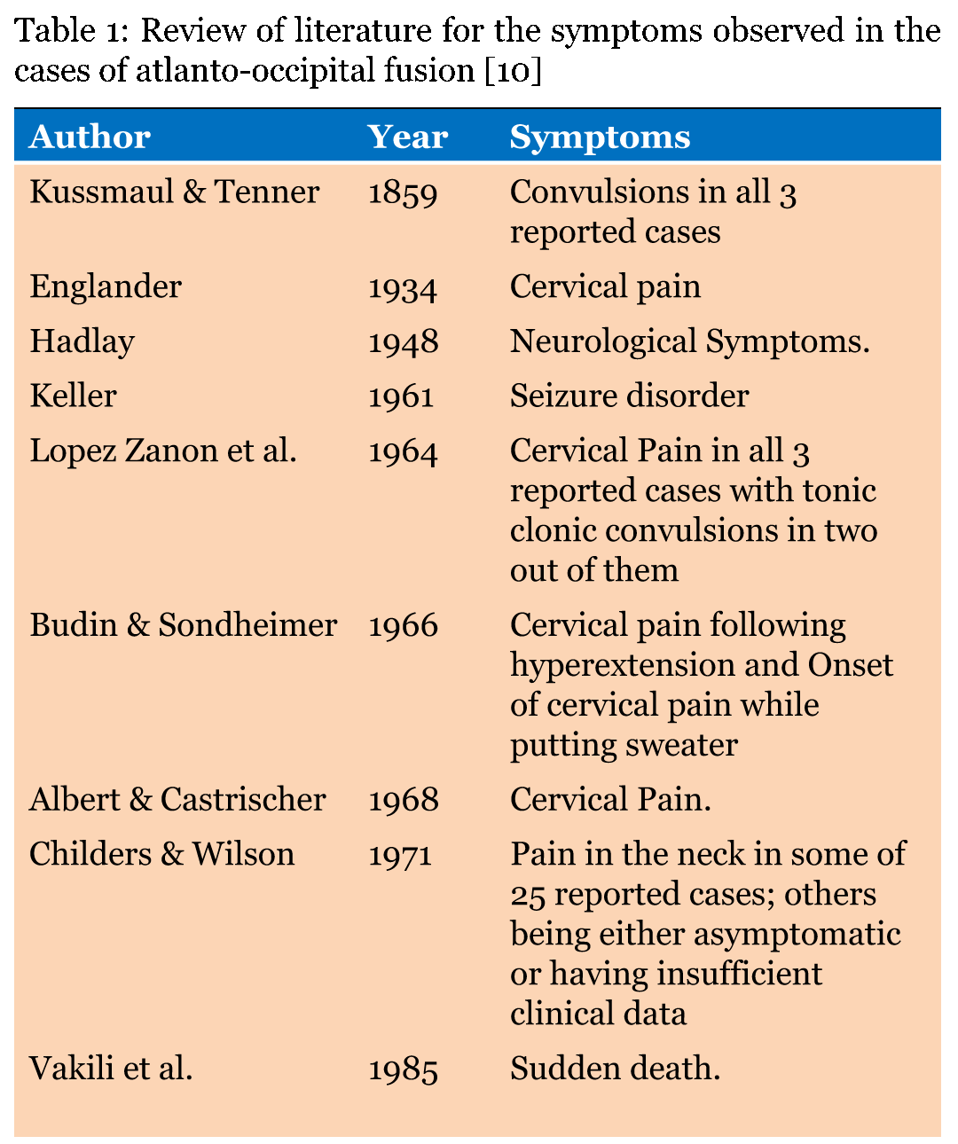
A skull with the atlanto-occipital fusion was noted during demonstration of norma basalis to first year MBBS students. So, to look for the similar finding in other skulls, total 50 human adult dry skulls were examined. No other skull had shown similar findings. The incidence of this anomaly in our study is 2%, i.e., one out of 50 skulls. In this skull with atlanto-occipital fusion, the left half of the anterior arch of the atlas vertebra is completely fused with the occipital bone of the skull. While the right half of the anterior arch is fused in the region of joint between occipital condyle and superior articular facet of atlas. On the posterior arch two halves of the arch fails to fuse in midline and forms the bifid arch. Both the halves of the posterior arch are fused with the margins of the foramen magnum. Transverse process of the right side is normal and not fused to the occipital bone but the transverse process of left side is broken. The inferior articular facet of the right side is normal but that on the left side articular facet is smaller in diameter, elongated and extends over the inferior aspect of the anterior arch (Figure 1). Transverse and antero-posterior diameter of the vertebral foramen of the atlas and foramen magnum appears normal. No other craniofacial abnormality was detected in this skull. | ||||||
| ||||||
|
Discussion
| ||||||
|
As quoted by Campos et al. [6] atlanto-occipital fusion was first described by Rokitansky in 1844 and demonstrated roentgenographically by Schuller in 1911. In humans, the craniovertebral junction corresponds to the boundary between fourth and fifth somites, but according to some authors it may be between fifth and sixth somites. Vertebrae are formed from the fifth somite caudally. Second, third and fourth occipital somites fused to form basiocciput. The first cervical vertebra is formed by the caudal half of fourth occipital somite and the cranial half of the first cervical somite. But the occipital condyles develop from the first cervical somite. In occipitocervical junctional region, the centra of the fifth, sixth, and seventh sclerotome has different fate than other caudally placed sclerotomes. The development of cervical spine, particularly the upper cervical vertebrae is closely related to the development of the basiocciput and exocciput. Anomalies of the occiput are associated with decrease skull base height, dense lies at or above the level of foramen magnum, margin of foramen magnum lies above the bottom of posterior cranial fossa, posterior arch of atlas is at the same level as the foramen magnum. Malfusion of the caudal portion of occipital sclerotome 4 and cranial portion of cervical sclerotome 1 may produce defects of the occipital condyles. Arnold-Chiari malformation is although consider clinically to be a neurological defect in which the medulla oblongata and sometimes the cerebellar tonsils project through the foramen magnum. There is good evidence that the underlying cause of this malformation is a series of abnormalities of occipitocervical junction. Atlas assimilation is found in this type of malformation [1]. As also quoted by Skartz et al. [7]failure of segmentation of the basal occipital sclerotome and first spinal sclerotome lead to osseous fusion between the atlas and the occipital bone. Cases of partial and complete fusion of atlas with occipital bone were found by several authors. Bose and Shrivastava [8] found the case of partial fusion of incomplete bony ring of the atlas to the base of occipital bone with different circumference of foramen magnum and vertebral foramen producing narrowing of foramen magnum, total fusion of lateral masses, anterior and posterior arches partially fused on left side, completely fused on right side, a separate foramen found on right side possibly for transmission of vertebral artery and first cervical nerve, asymmetry in size of inferior articular facets. In such cases, the chances of neurological complications are more due to compression of spinal cord and other nerves and vessels like vertebral arteries. Rajani et al. found a case of unilateral fusion of left half of atlas with corresponding occipital bone [9]. Skull showed asymmetries on left side like change in size of neurovascular foramina, shifting of position of styloid process, reduction in size of middle cranial fossa along with corresponding petrous bone. Campos et al. found a case of atlanto-occipital fusion in which the posterior arch of the atlas vertebra showed bifid form and there was the absence of the foramen of the transverse process on both sides of the atlas vertebra [6]. As quoted in literature clinical symptoms in cases of atlanto-occipital fusion varies from simple cervical pain to sudden death (Table 1) [10]. Incidence of the atlanto-occipital fusion is less but considering the symptomatology this variation is important to be aware of (Table 2). | ||||||
| ||||||
| ||||||
|
| ||||||
|
Conclusion
| ||||||
|
Atlanto-occipital fusion is a rare anomaly which requires awareness among neurosurgeons, physicians, and orthopedicians due to its common clinical symptoms but dire neurological consequences like sudden death. | ||||||
|
References
| ||||||
| ||||||
|
[HTML Abstract]
[PDF Full Text]
|
|
Author Contributions
Dipti A. Nimje – Substantial contributions to conception and design, Drafting the article, Revising it critically for important intellectual content, Final approval of the version to be published Harish A. Wankhede – Substantial contributions to conception and design, Drafting the article, Revising it critically for important intellectual content, Final approval of the version to be published |
|
Guarantor of submission
The corresponding author is the guarantor of submission. |
|
Source of support
None |
|
Conflict of interest
Authors declare no conflict of interest. |
|
Copyright
© 2014 Dipti A. Nimje et al. This article is distributed under the terms of Creative Commons Attribution License which permits unrestricted use, distribution and reproduction in any medium provided the original author(s) and original publisher are properly credited. Please see the copyright policy on the journal website for more information. |
|
|
|
About The Authors
| |||
| |||
| |||