| Table of Contents | |
|
Original Article
| ||||||
| Using the balance between proliferation and apoptosis to assess the cryopreservation and thawing protocol in mouse 4-cell embryos | ||||||
| Mostafa M. El-Naggar1, Hassan Nasrat2, Hassan Jamal2, Samar Al-Saggaf3, Mohamed H. Badawod3 | ||||||
|
1Professor, Department of Anatomy, Faculty of Medicine, Jazan University, Jazan, Saudi Arabia.
2Professor, Department of Obstetrics and Gynecology, Faculty of Medicine, King Abdulaziz University, Jeddah, Saudi Arabia. 3Professor, Department of Anatomy, Faculty of Medicine, King Abdulaziz University, Jeddah, Saudi Arabia. | ||||||
| ||||||
|
[HTML Abstract]
[PDF Full Text]
[Print This Article]
[Similar article in Pumed] [Similar article in Google Scholar] |
| How to cite this article |
| El-Naggar MM, Nasrat H, Jamal H, Al-Saggaf S, Badawod MH. Using the balance between proliferation and apoptosis to assess the cryopreservation and thawing protocol in mouse 4-cell embryos. Edorium J Anat Embryo 2015;2:6–13. |
|
Abstract
|
|
Aims:
The criteria used to assess the optimal conditions for cryopreservation of the embryos in the in vitro fertilization (IVF) protocols are still a matter of discussion. This study aimed at evaluating the use of cell proliferation and apoptosis to assess the optimal conditions for cryopreservation/thawing of the 4-cell embryos.
Methods: Fertilized ova were collected from mated female MF1 mice 24 hours after hCG injection. They were cultured in KSOM medium and kept in CO2 incubator at 37°C and 5% CO2 up to the stage of the 4-cells. Two methods of cryopreservation were used; the step-rate and the ultra-rapid vitrification. Slow and fast thawing was done. Slides were prepared from samples of the embryos, and stained immunohistochemically for proliferative and apoptotic cells. The proliferative capacity was measured by labeling with bromodeoxyuridine (BrdU) and the apoptotic ability was measured with TUNEL technique. Results: Vitrification with fast thawing of the 4-cell embryos gave better morphology, higher proliferative capacity, and lower apoptotic ability. Following step-rate cryopreservation with slow or fast thawing, cell labeling index for BrdU was 0% and 17%, respectively and was 66% and 83%, respectively following vitrification. The incidence of apoptosis following step rate cryopreservation with slow or fast thawing was 96% and 89%, respectively and was 42% and 13%, respectively following vitrification. Conclusion: It is concluded that cell proliferation and apoptosis could be used to assess the cryopreservation/thawing protocol for early embryos. | |
|
Keywords:
Apoptosis, Cryopreservation, Embryos, Mouse, Proliferation, Thawing
| |
|
Introduction
| ||||||
|
In vitro fertilization (IVF) has become an established procedure to treat infertility. Cryopreservation of the extra embryos at –196°C would allow repeating the IVF procedure with the advantage of avoiding the inconvenient induction of ovulation and the invasive procedure of oocyte retrieval. It would also give flexibility to the IVF program and allow selecting the proper time for the recipient mother for receiving the embryos. The specimens are stored at –196°C, where at this low temperature, storage for long periods theoretically have minimal impact on viability [1]. The optimal conditions for cryopreservation/thawing protocol for the early embryos are still a matter for discussion. Various criteria were used to assess the optimal conditions for cryopreservation of the embryos; such as in vitro light and electron microscopic morphological changes [2] [3] [4] [5] , metabolic activity [6], in vitro and in vivo embryo development [2] [3] [7][8]. Since growth of the embryo is supported by increased cell proliferation and low apoptosis, these two factors are critical for further development of the embryo. However, the effect of cryopreservation on the proliferation/ apoptosis equilibrium of the embryonic cells was not investigated. The present work aimed at using proliferative capacity and the apoptotic ability of the embryonic cells to evaluate different cryopreservation/ thawing protocols of the 4-cell embryos. The proliferative capacity was measured by labeling the proliferating cells with BrdU. The apoptotic ability was measured by labeling the apoptotic cells with the TUNEL technique. Morphological changes were also used for comparison. Two cryopreservation methods (step-rate and ultra-rapid vitrification), and two thawing rates (slow and fast) were used. | ||||||
|
Materials and Methods
| ||||||
|
Collection of fertilized ova Cryopreservation/thawing The cryopreserved embryos were thawed by the slow or fast warming rates. For slow thaw the cryotubes were kept standing on the bench at room temperature for approximately 10 min. For fast-thaw the cryotubes were agitated in a water bath at 37°C. In both cases the tubes were transferred to an ice bath at 0°C, just before lyses of the last ice crystal. DMSO was drawn from the intracellular compartment by a hyperosmolar sucrose solution, which was then gradually diluted and replaced by culture medium. The ultra-rapid (vitrification) procedure is the method described by Kasai et al. (1990) [1]. The procedure consisted of washing the embryos in Dulbeco's phosphate buffered saline (D-PBS). Embryos were equilibrated in the vitrification solution for two minutes to dehydrate the cytoplasm. The vitrification solution (EFS40) consisted of 40% ethylene glycol in solution of 30% Ficoll, 0.5 M sucrose and BSA dissolved in D-PBS. The embryos were then transferred to 13 mm EFS40 column in the straw. The straw was allowed to cool slowly in liquid nitrogen vapor for at least three minutes before immersing in liquid nitrogen (-196°C) for storage. The vitrified embryos were thawed by the slow or fast warming rates. For slow thaw, the straws were kept standing on air at room temperature for 15 seconds, and then immersed into a 20°C water bath. For fast thaw, the straws were agitated in a water bath at 37°C. When the sucrose solution began to melt, the straws were removed from the water bath and slowly perfused with 1 ml sucrose solution, the embryos recovered and transferred to drops of hyperosmolar sucrose in culture dish. Glucose was then gradually diluted and replaced by culture medium. Morphological examination Immunohistochemical examination BrdU labeling for proliferation TUNEL technique for apoptosis TUNEL Technique was used to measure programmed cell death (apoptosis) by detecting DNA strand breaks in individual cells. It uses terminal deoxy-transferase (TdT) to label free 3'OH ends in genomic DNA with fluorescein-dUTP. The slides were incubated with TUNEL reaction mixture containing TdT and fluorescein-dUTP. During this incubation step, TdT catalyzes the attachment of fluorescein-dUTP to the free 3'OH ends in the DNA. The incorporated fluorescein is detected with an anti-fluorescein antibody peroxidase conjugate. The immune complex peroxidase is then visualized by a substrate reaction and detected by light microscope. The negatively stained nuclei were counter-stained with hematoxylin. The cell labeling index (percentage of cells labeled) was measured by counting the number of TUNEL labeled nuclei and express it as a percentage of the total number of nuclei scored. Statistical Analysis The significance of difference between the cryopreserved and control specimens was evaluated by students' t-test at p < 0.05. | ||||||
|
Results | ||||||
|
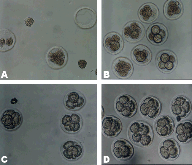
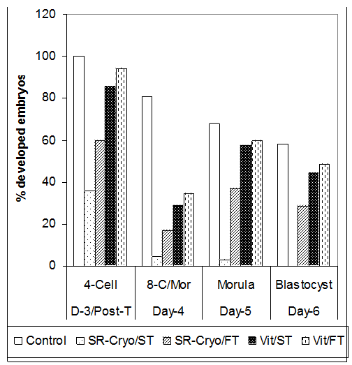
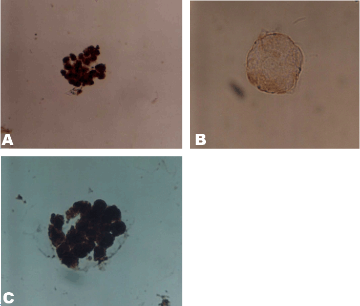
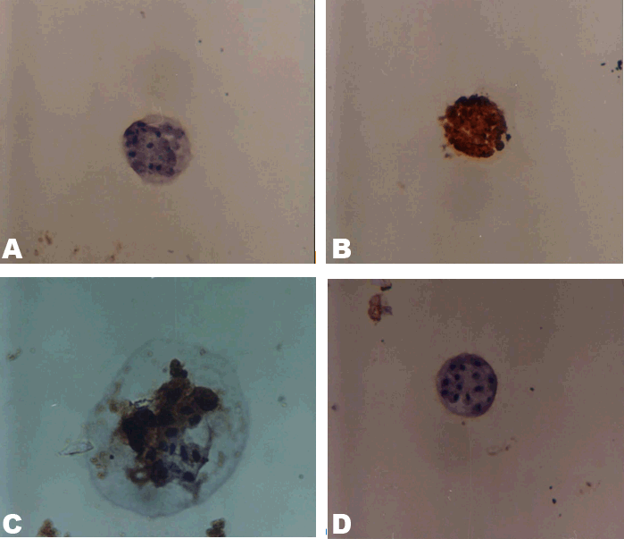
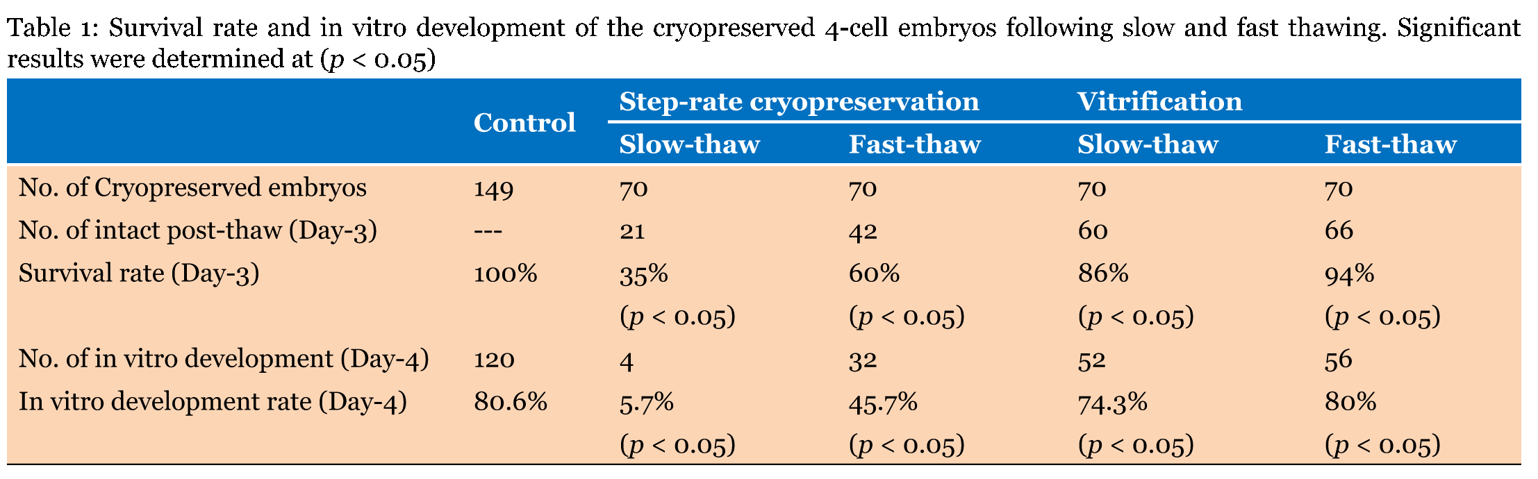
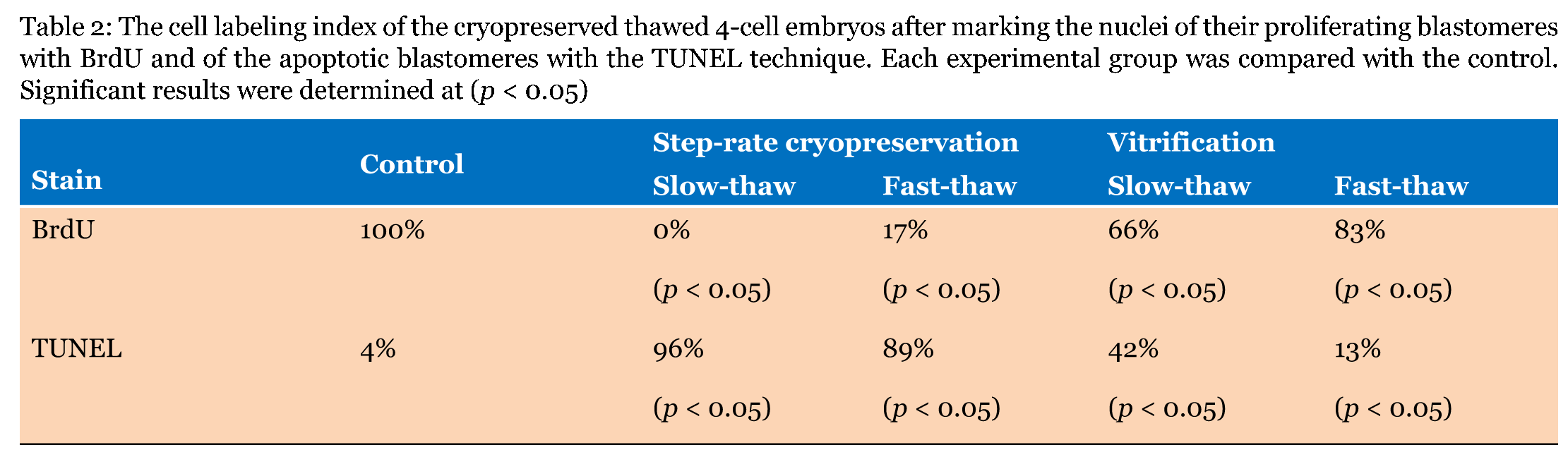
Morphological findings Vitrified 4-cell embryos with slow-thawing appeared in good condition with spherical well-defined borders of the blastomeres surrounded with thick zonae (Figure 1C). Similar results were found in the fast-thawed embryos which showed spherical thick zona pellucida with four well defined blastomeres inside (Figure 1D). The step-rate cryopreserved embryos showed survival rates of 35% and 60%, and in vitro development rates of 6% and 46% with slow and fast thawing, respectively (Table 1). The best results were obtained with vitrification of the embryos both with slow and fast thawing, which gave survival rates of 86% and 94%, and in vitro development rates of 74% and 80%, respectively (Table 1). The in vitro differential developmental progress of the cryopreserved/ thawed embryos was much slower than that of the corresponding control embryos, especially at day-4 (stage of 8-cell mass/ morula). During day-5 (stage of morula) and day-6 (stage of blastocyst), their developmental progress was slightly slower than that of the control (Figure 2). Bromodeoxyuridine (BrdU) labeling for proliferation The cell labeling index (percentage of cells labeled) for BrdU was nearly 100% in the control group. Following step-rate cryopreservation and slow or fast thawing, cell labeling index was 0% and 17%, respectively. Following vitrification and slow or fast thawing, cell labeling index was 66% and 83%, respectively (Table 2). TUNEL technique for apoptosis The cell labeling index (percentage of cells labeled) for TUNEL showed that the incidence of apoptosis in the control embryos is about 4%. The incidence of apoptosis has increased following step rate cryopreservation and vitrification to 96% and 42%, respectively after slow thawing and to 89% and 13 %, respectively after fast thawing (Table 2). | ||||||
|
| ||||||
|
| ||||||
|
| ||||||
|
| ||||||
| ||||||
|
| ||||||
|
| ||||||
|
Discussion
| ||||||
|
The results of this study showed that vitrification with fast thawing gave the best proliferation cell labeling index (83%) and the least incidence of apoptosis (13%). These proliferation/apoptosis results were more or less supportive to the morphological evaluation; survival rate of 94% and in vitro development rate of 80% following vitrification and fast thawing. The results of the morphological study obtained in this report are comparable to those obtained by other authors. The recorded survival rate following vitrification methods was 83–94% [11] in mouse, 63% [12], and 79.2% [13] in human embryos. High rates of in vitro development following vitrification was reported to be 84–98 % [1] in rat, 75–97% [14], 80–83% [15], 74–92% [16], 94–95% [17] in mouse, and 80% [18] in human embryos. Although we could not find in literature any report critically investigating the effect of cryopreservation on both proliferation and apoptosis of the early embryonic cells, however, few reports could be located investigating the effect of either proliferation or apoptosis. Few investigations used proliferation ratio to provide information about the developmental potential of embryos after cryopreservation [19] [20] . Takagi et al. (1996) [21] investigated proliferation of the inner cell mass (ICM) by examining the rate of DNA synthesis in frozen-thawed bovine blastocysts, by immunocytochemical staining. They found that the numbers of bromodeoxyuridine- immunoreactive ICM cells of frozen-thawed embryos were significantly lower than those of unfrozen embryos, suggesting that the rates of proliferation of ICM of frozen-thawed bovine embryos tend to be lower than those of unfrozen embryos. Few reports used the dUTP nick end-labeling (TUNEL) technique to describe the effect of cryopreservation on the apoptotic cell death [5] [22] [23] [24] [25] [26]. The mechanisms underlying the optimum control of the proliferation/apoptosis balance for embryonic development following in vitro fertilization and culture are unknown. Several factors have been investigated and claimed responsible for increasing or decreasing proliferation of early embryos. It has been suggested that lipid peroxides derived from polyunsaturated fatty acids (PUFAs) inhibit proliferation of various cells, a process that could be reversed by antioxidants [27]. Several factors could be involved in early embryonic cellular proliferation and development; such as Brca1 [28], tumor suppressor gene Brca2 [29] , radiation-inducible gene muREC2/RAD51L1 [30], insulin [31], cyclin proteins [32], embryo-derived platelet-activating factor (EPAF) [33], polyamines [34], and all-trans retinoic acid [35]. Apoptosis, on the other hand, was found to be affected by several factors [36] [37][38]. Levy et al. (1998) [39] has shown that apoptosis occurs in mammalian embryos as early as the cleavage stage, in order to regulate the inner cell mass. Current evidence indicates that hydrogen peroxide causes apoptosis of inner cell mass cells destined to develop into trophectoderm, the first apoptotic event during mammalian development [37] [40]. Yang and Rajamahendran (2002) [38] showed that apoptosis appears to be due to an interactions between the Bcl-2 family of proteins in pre-implantation embryos development. | ||||||
|
Conclusion
| ||||||
|
It is concluded that proliferative capacity and apoptotic ability of the embryonic cells could be used as criteria for assessing cryopreservation/thawing protocols. Disturbance of this balance, in the form of inhibited cellular proliferation or stimulated apoptosis, may compromise the developmental process of the growing embryos. It is possible that these two processes were minimally disturbed with vitrification and fast thawing of the 4-cell embryos, since this procedure gave the best development rate in vitro. These results may have an impact on the human IVF protocol and developing a bank of embryos that could be cryopreserved for future use. | ||||||
|
Acknowledgements
| ||||||
|
This work was supported by grant no. 003/420 from King Abdulaziz University, Jeddah, Kingdom of Saudi Arabia. | ||||||
|
References
| ||||||
| ||||||
|
[HTML Abstract]
[PDF Full Text]
|
|
Author Contributions:
Mostafa M. El-Naggar – Substantial contributions to conception and design, Acquisition of data, Analysis and interpretation of data, Drafting the article, Revising it critically for important intellectual content, Final approval of the version to be published Hassan Nasrat – Substantial contributions to conception and design, Acquisition of data, Analysis and interpretation of data, Drafting the article, Revising it critically for important intellectual content, Final approval of the version to be published Hassan Jamal – Substantial contributions to conception and design, Acquisition of data, Analysis and interpretation of data, Drafting the article, Revising it critically for important intellectual content, Final approval of the version to be published Samar Al-Saggaf – Substantial contributions to conception and design, Acquisition of data, Analysis and interpretation of data, Drafting the article, Revising it critically for important intellectual content, Final approval of the version to be published Mohamed H. Badawod – Substantial contributions to conception and design, Acquisition of data, Analysis and interpretation of data, Drafting the article, Revising it critically for important intellectual content, Final approval of the version to be published |
|
Guarantor of submission
The corresponding author is the guarantor of submission. |
|
Source of support
None |
|
Conflict of interest
Authors declare no conflict of interest. |
|
Copyright
© 2015 Mostafa M. El-Naggar et al. This article is distributed under the terms of Creative Commons Attribution License which permits unrestricted use, distribution and reproduction in any medium provided the original author(s) and original publisher are properly credited. Please see the copyright policy on the journal website for more information. |
|
|