| Table of Contents | |
|
Case Report
| ||||||
| Triple ectopic thyroid associated with normally located pretracheal thyroid gland: A rare entity | ||||||
| Gabriel J. Mchonde1, Hortensia G. Nondoli2, Masato Hirakawa3 | ||||||
|
1PhD, Lecturer, Department of Biomedical Sciences, School of Medicine and Dentistry, College of Health Sciences, University of Dodoma, Dodoma-Tanzania.
2MSc, Lecturer, Department of Anatomy and Histology, School of Medicine, University of Dar Es Salaam, Dar Es Salaam-Tanzania. 3MSc, Assistant Lecturer, Department of Cell Biology and Anatomy, School of Medicine, Iwate Medical University, Iwate-Japan. | ||||||
| ||||||
|
[HTML Abstract]
[PDF Full Text]
[Print This Article]
[Similar article in Pumed] [Similar article in Google Scholar] |
| How to cite this article |
| Mchonde GJ, Nondoli HG, Hirakawa M. Triple ectopic thyroid associated with normally located pretracheal thyroid gland: A rare entity. Edorium J Anat Embryo 2016;3:34–38. |
|
Abstract
|
|
Introduction:
Triple ectopic thyroid tissues associated with a normally located thyroid is a rare encountered anomaly among thyroid dysgenesis.
Case Report: Herein, we present a case observed during a routine dissection on a 88-year-old embalmed female cadaver who had a normally located pretracheal thyroid gland co-existed with three independent inferiorly located thyroid tissue masses extended into the superior mediastinum displacing the aortic arch and associated major vessels. Conclusion: Thyroid dysgenesis can be genetic and inheritable due to involvement of mutations on regulatory genes required for early embryonic development of the thyroid gland. More information on these rare anatomical variations could benefit physicians, surgeons and radiologists on differential diagnosis and management strategies during their routine procedures. | |
|
Keywords:
Cervical, Superior mediastinum, Thyroid anomalies, Triple ectopia
| |
|
Introduction
|
|
Morphogenesis disturbances on establishment, migration and organization of thyroid primordium occurring during early stages of embryogenesis may results to generation of ectopic thyroid glands or ectopic thyroid tissues along the gland's embryological descending pathway from foramen cecum to the mediastinum, as well as distant areas [1] [2] [3]. Ectopic thyroid is formed when the thyroid primordium or a portion of it fails to descend along the normal migration pathway and results into the gland development in an anomalous position. Multiple ectopic foci of thyroid gland (tissues) are very rare and only few cases of dual and triple ectopic have been reported with frequent locations along the midline from base of the tongue to the mediastinum or sub diaphragmatic organs [4] [5] [6]. However, most of the reported cases are reporting the incidence of multiple ectopic being in various locations on the cervical region above or lateral to the normally located thyroid gland, or thyroid tissues embedded within other organs. Here, we are reporting a case of triple ectopic thyroid located inferior to the normally located thyroid gland and extending into superior mediastinum between great vessels and the trachea. |
|
Case Report
|
|
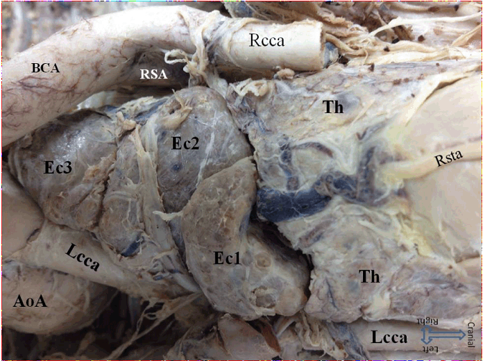
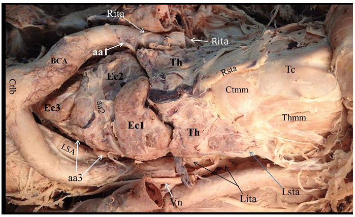
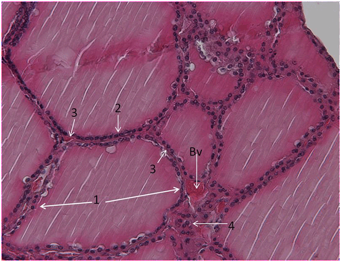
During routine head and neck dissection classes for the undergraduate medical students, we encountered variation on number and position of the thyroid gland of a 88-years-old formalin embalmed female cadaver. After the anterior cervical and upper thoracic regions skin and superficial and deep fascia were removed, we encountered a normally positioned pretracheal thyroid gland associated with three other inferiorly placed ectopic thyroid masses of which each had its own blood supply and venous drainage, and extending from the cervical to superior mediastinum displacing the aortic arch and great vessels anteriorly, and trachea laterally to the right. Normal thyroid gland was located anterior to the trachea, inferior to the thyroid cartilage of the larynx and had a typical butterfly-shape appearance with two lobes (left and right) connected by an isthmus and extends inferiorly to fifth tracheal ring. Each lobe measured 4.5 cm long, 2.8 cm wide, 1.5 cm thick, and received blood supply from superior and inferior thyroid arteries originated from external carotid and common carotid arteries respectively (Figure 1) and (Figure 2). Ectopic-thyroid-I was a tongue-shaped extending from left to right located inferior to the normally positioned thyroid gland and superior to the ectopic-thyroid-II. It measured 2.2 cm long, 4 cm wide with 2 cm thick at the broad end, and 1cm at its tapered end (Figure 2). It received blood supply from artery branched from the left inferior thyroid artery originating from left common carotid artery. Ectopic-thyroid-II was triangular in shape bordered superiorly by normal thyroid and ectopic-thyroid-II, and ectopic-thyroid-III inferiorly and extended into superior mediastinum displacing laterally the carotid and subclavian arteries. It measured 2.4 cm thick, 4 cm on its longest side, 3 cm and 2.8 cm on other sides (Figure 2). It had dual blood supply from right common carotid artery and left inferior thyroid artery. Ectopic-thyroid-III was a triangular in shape with its apex extending deep into the superior mediastinum intertwined between the aortic arch (anterior) and trachea (posterior) at the level of thoracic vertebral IV (Figure 1) and (Figure 2)and displacing the aortic arch anteriorly and the trachea posterolateral to the right. It received dual blood supply from right common carotid artery and a branch from left inferior thyroid artery. Associated variations Histology |
|
|
|
|
|
|
|
Discussion
|
|
Thyroid ectopia are rarely encountered developmental anomalies although it is the most frequent form of thyroid dysgenesis [3] [7] and its presence may be symptomatic or asymptomatic clinically depending on location, size and functional activities. Dual thyroid ectopia has been reported to associate lingual and submandibular thyroid or intranasal and nasopharynx thyroid ectopia co-existing with normally located thyroid gland [5] [6]. Triple ectopic have been reported to involve lateral ectopic thyroid masses co-exist with eutopic thyroid gland, or sublingual-suprahyoid-subhyoid thyroids [4] or foramen cecum-suprahyoid-cricoid thyroids or lingual-hyoid-suprahyoid thyroids, all with the absence of normally located thyroid gland. However, altogether these cases are reporting the ectopic being on the lateral or superior to the normally located thyroid gland, and none of them are reporting three distinct independent thyroid masses located inferior to the normally located thyroid gland. Intrathoracic thyroid ectopia has been also reported previously in the mediastinum, lungs, and heart and mostly were discovered incidentally on chest radiograph or during autopsy. However, the reported mediastinal thyroid ectopia were located in the anterior or posterior mediastinum [8] and none in the superior mediastinum as observed in the current case displacing the aortic arch, great vessels and the trachea. Embryologically, the thyroid primordium develops about 24th day post-fertilization as median endodermal thickening in the floor of primitive hypopharynx which then descend along the migration pathway from foramen caecum towards its normal position. Definitive thyroid gland cells develop as a result of merging of migrating cells from thyroid anlage (follicular cells) and ultimobranchial bodies (parafollicular cells) from fourth pharyngeal pouch. Ectopic thyroid tissues may rise as a faulty embryogenesis which could be due to mutation of thyroid transcription factors required for descending migration [3][4] or developmental defects other than abnormal migration of the thyroid primordium. Genetic defects involving mutations in the paired box transcription factors PAX8 and thyroid transcription factors TTF1 and TTF2 has been linked with thyroid dysgenesis [3] [9]. Mediastinal ectopia or other anomaly position could be due displacement of the mesenchymal tissue during the course of embryonic development. In the current case, three independent ectopic thyroids located inferior to the normally located thyroid but along the midline of the neck to the superior mediastinum between trachea and aortic arch were observed. Conceivable clarifications for this entity could be due to the division of the thyroid primordium caused by the developing trachea and its cartilage rings. The condition may also be hypothesized to arise as a result of aberrant migration and differentiation of uncommitted endodermal cells of the thyroid primordium along the midline. |
|
Conclusion
|
|
Alterations in signaling or defects during early embryogenesis of thyroid gland generate thyroid ectopia along its embryological pathway or distant areas. Multiple ectopic thyroid tissue are rare developmental defects and could be difficult to diagnose since most cases are asymptomatic. In the present case, three independent ectopic thyroid tissues extend from the neck inferior to the normally positioned thyroid gland to the superior mediastinum could be due to the developmental defects other than migration defects such as division of the developing thyroid gland caused by trachea and its cartilage rings or by aberrant migration and differentiation of uncommitted endodermal cells from the thyroid primordium. Understanding of this rare entity could benefit the physician, surgeons and radiologists during their routine procedures performed within or closely to the superior mediastinum and also in differentiating the mediastinal masses. |
|
Acknowledgements
|
|
We expressing thanks to Prof. Yoh-ichi Satoh and Prof. Sumio Isogai for their moral and material support and all members in the Department of Anatomy, Iwate Medical University for their encouragement. |
|
References
|
|
|
Suggested Reading
|
|
|
[HTML Abstract]
[PDF Full Text]
|
|
Author Contributions
Gabriel J. Mchonde – Substantial contributions to conception and design, Acquisition of data, Analysis and interpretation of data, Drafting the article, Critical revision of the article, Final approval of the version to be published Hortensia Nondoli G. – Analysis and interpretation of data, Critical revision of the article, Final approval of the version to be published Masato Hirakawa – Acquisition of data, Analysis and interpretation of data, Final approval of the version to be published |
|
Guarantor of submission
The corresponding author is the guarantor of submission. |
|
Source of support
None |
|
Conflict of interest
Authors declare no conflict of interest. |
|
Copyright
© 2016 Gabriel J. Mchonde et al. This article is distributed under the terms of Creative Commons Attribution License which permits unrestricted use, distribution and reproduction in any medium provided the original author(s) and original publisher are properly credited. Please see the copyright policy on the journal website for more information. |
|
|