| Table of Contents | |
|
Letters to the Editor
| ||||||
| The genetics in sulci and gyri development | ||||||
| Andrew J. Flocchini | ||||||
|
17050 Old Lakeville Road 3, Petaluma, CA.
| ||||||
| ||||||
|
[HTML Abstract]
[PDF Full Text]
[Print This Article]
[Similar article in Pumed] [Similar article in Google Scholar] |
| How to cite this article |
| Flocchini AJ. The genetics in sulci and gyri development. Edorium J Anat Embryo 2016;3:51–53. |
|
To the Editors,
| ||||||
|
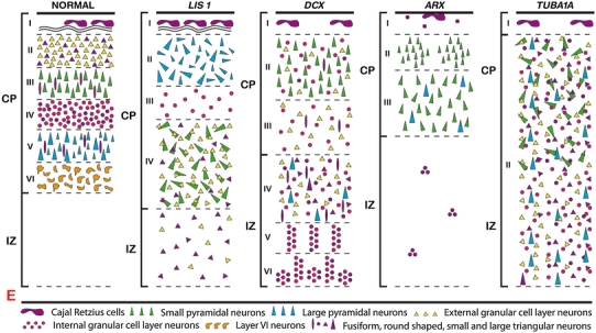
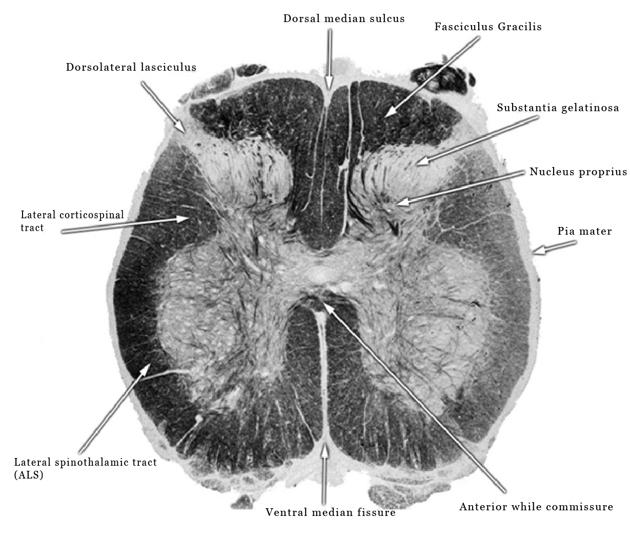
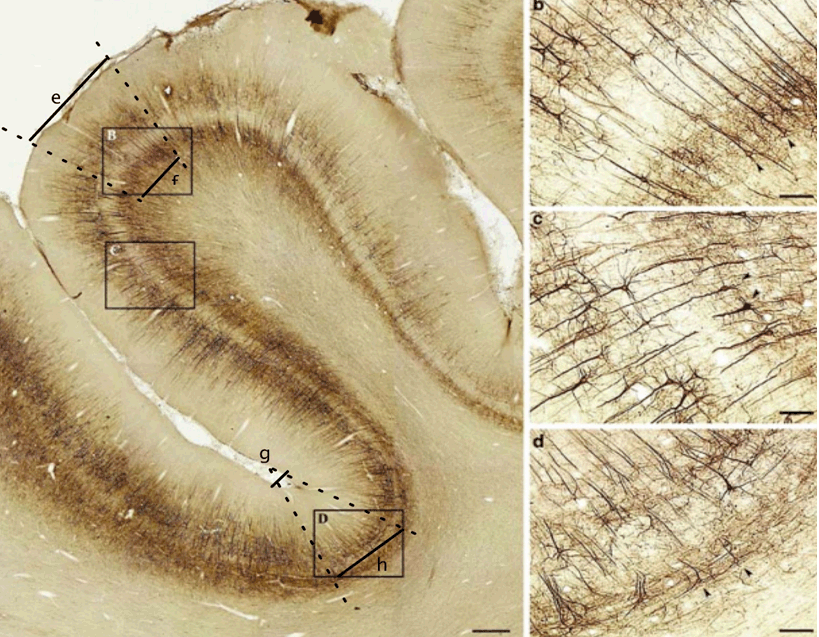
Sulci and gyri are the ridges and valleys on the surface of the brain. How these develop have been studied for decades without much success. Most studies show that buckling forms sulci and gyri by the force and compression of cell layers inside the brain [1] [2][3][4], but compression could affect the dendrite branches and axon terminals of neurons and their connections. Knowing how a particular development takes place and where this development is abnormal may help show other disorders of the brain such as lissencephaly (Figure 1). Here I show how the meninges may form sulci and gyri in the Rhesus monkey brain. In this manner, I theorize that the formation of the cortex is done by an outside pull, as compared to compression and force from the inside. The meningeal tissue is the same from the surface of the brain to the tip of the spinal cord [5]. The dorsal median sulcus and the ventral median fissure of the spinal cord, (Figure 2), show a strong genetic influence having the same blueprint structure in all humans. If the notion that genetic influence of the meningeal tissue on the spinal cord is correct, this could also be applied to the central and lateral sulci in the brain, which are seen in all humans, and they are also connected to all other sulci and gyri (Figure 3) rhesus monkey brain. In the gyrus, there is a higher ratio of pial and arachnoid mater to neurons which have more elongated axons as compared to the lower ratio in the sulcus which have more disturbed axons as seen in 'd-arrows'. The higher magnification image of 'c' has a 1 to 1 ratio of pia mater to neurons showing a slight side pull of axons to the gyri above. The distance between the neurons and the pia mater in images 'b' and 'c' are the same, suggesting that neuronal distance from the pia mater is not a factor in the difference of axonal form. As one can see in Figure 3, the gyrus is wider than the sulci on either side, giving the appearance that it has been pulled or vacuumed by an outside force. The formation of sulci may begin by the arachnoid mater detaching from the pia mater weakening the neuronal attraction causing the thin flat cells of the pia mater to buckle creating the gyri. The meningeal tissue of the future sulcus may be static while gyri form on either side. The mechanism in the pial tissue that causes the axons to become elongated could also be the mechanism that causes the skull to grow in relation to the brain. Current studies suggest that skull growth is dictated by the brain size [6]. However, the mechanism in the pia mater that causes elongated neurons could also be the mechanism that causes the activation of osteoclasts and the destruction of the endocranium, while simultaneously creating stimuli to activate osteoblasts on the ectocranial surface to create new bone. This process of simultaneous destruction and creation of skull bone can be seen as a push-pull relationship between the brain and the skull. | ||||||
| ||||||
| ||||||
|
| ||||||
|
| ||||||
|
Conclusion
| ||||||
|
The process of how sulci and gyri form in the brain has been studied for years without much success. Knowing how a developmental process takes place can help explain the causes of developmental disorders, such as lissencephaly. The axonal form is very similar to the axonal form found in lissencephaly. If the meningeal tissue is what controls axonal form, then the absence of arachnoid mater in sulci could cause a decrease in the strength of the signal that controls axonal form. An overall lesser signal throughout the meningeal tissue, as in a brain with lissencephaly, could explain why axonal form is highly disturbed throughout. Keywords: Brain, Genetics, Sulci and gyri | ||||||
|
Acknowledgements
| ||||||
|
Jordan James for editing the paper | ||||||
|
References
| ||||||
| ||||||
|
[HTML Abstract]
[PDF Full Text]
|
|
Author Contributions:
Andrew J. Flocchini – Substantial contributions to conception and design, Acquisition of data, Analysis and interpretation of data, Drafting the article, Revising it critically for important intellectual content, Final approval of the version to be published |
|
Guarantor of submission
The corresponding author is the guarantor of submission. |
|
Source of support
None |
|
Conflict of interest
Authors declare no conflict of interest. |
|
Copyright
2016 Andrew J. Flocchini. This article is distributed under the terms of Creative Commons Attribution License which permits unrestricted use, distribution and reproduction in any medium provided the original author(s) and original publisher are properly credited. Please see the copyright policy on the journal website for more information.
|
|
|