|
Clinical Image
“Eyes at the back head,” Incidental finding of giant parietal foramina: Foramina parietalia permagna
1 MD, Radiology, Hospital University Fann, Dakar, Senegal
2 MD, Anatomy Lab, University of Thies, Thies, Senegal
3 Pr, Radiology, Hospital University Fann, Dakar, Senegal
Address correspondence to:
Ibrahima NIANG
Avenue Cheikh Anta DIOP, BP 5035, Dakar,
Senegal
Message to Corresponding Author
Article ID: 100028A04IN2021
Access full text article on other devices

Access PDF of article on other devices


How to cite this article
NIANG I, NDAO CL, MAR NB, BA S. “Eyes at the back head,” Incidental finding of giant parietal foramina: Foramina parietalia permagna. Edorium J Anat Embryo 2021;8:100028A04IN2021.ABSTRACT
No Abstract
Keywords: Foramina parietalia permagna, Parietal bone, Parietal foramina
Case Report
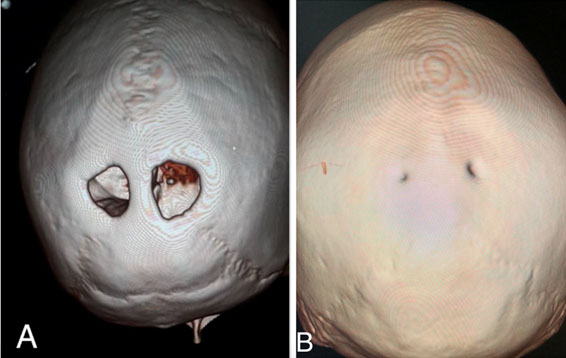
We report the case of a 28-year-old male patient with no specific surgical or medical history, who consulted for subacute headaches without other associated clinical signs.
A brain computed tomography (CT) scan was performed in this patient. This contrast CT scan did not reveal any cerebral, ventricular, vascular, or other cerebral lesion.
Exploration of the skull in bone window and in 3D made it possible to visualize a huge bilateral and symmetrical dilation of the parietal foramina on either side of the sagittal suture, measured at 2 × 2.5 cm to the right and 2.3 × 2.7 cm to the left, giving the appearance of “eyes at the back of the head’’ (Figure 1A) and corresponding to a foramina parietalia permagna. To highlight the extent of the dilatation, we show an image of a small parietal foramina in another patient (Figure 1B).
Discussion
“Foramina parietalia permagna,” also called “The Catlin Mark,” is a congenital anatomical variant with autosomal dominant inheritance. It is extremely rare, estimated 1 in 25,000 cases [1],[2]. Greig [3] considers it to be “sufficiently rare to make it desirable that every example be reported.”
It corresponds to a lack of ossification of the posterior membranous parietal bones during embryogenesis, leaving open parietal foramina, on each side of the sagittal suture, ranging in size from a few millimeters to approximately 2 cm.
It is most often benign, causing at most headaches and vomiting, although associations have been reported with other congenital anomalies such as cleft palate, myelomeningocele, encephaloceles, epilepsy and other defects.
Family transmission seems to be the rule, as first observed in 16 of the 56 members of the Catlin family by Goldsmith (1922) [1]. Further studies have proven the relation with heterozygous mutations of the genes ALX4 on chromosome 11 and MSX2 on chromosome 5 [4].
These bone defects could be mistaken for osteolysis, trauma (gunshot), or iatrogenic (craniotomy). They could also be useful in postmortem identification by the skull, as part of thanatoradiology or a conventional autopsy.
Conclusion
Foramina parietalia permagna is a rare congenital anatomical variant that deserves to be known and recognized, in particular by radiologists and forensic pathologists. It is most often benign but its discovery should lead to a search for other malformations that may be associated with it.
REFERENCES
1.
Goldsmith WM. “The Catlin mark” the inheritance of an unusual opening in the parietal bones. Journal of Heredity 1922;13(2):69–71.

2.
Griessenauer CJ, Veith P, Mortazavi MM, et al. Enlarged parietal foramina: A review of genetics, prognosis, radiology, and treatment. Childs Nerv Syst 2013;29(4):543–7. [CrossRef]
[Pubmed]
3.
Greig DM. Congenital and symmetrical perforation of both parietal bones. J Anat Physiol 1892;26(Pt 2):187–91.
[Pubmed]
4.
Mavrogiannis LA, Taylor IB, Davies SJ, Ramos FJ, Olivares JL, Wilkie AOM. Enlarged parietal foramina caused by mutations in the homeobox genes ALX4 and MSX2: From genotype to phenotype. Eur J Hum Genet 2006;14(2):151–8. [CrossRef]
[Pubmed]
SUPPORTING INFORMATION
Author Contributions
Ibrahima NIANG - Conception of the work, Design of the work, Acquisition of data, Analysis of data, Drafting the work, Final approval of the version to be published, Agree to be accountable for all aspects of the work in ensuring that questions related to the accuracy or integrity of any part of the work are appropriately investigated and resolved.
Coumba Laobé NDAO - Acquisition of data, Drafting the work, Final approval of the version to be published, Agree to be accountable for all aspects of the work in ensuring that questions related to the accuracy or integrity of any part of the work are appropriately investigated and resolved.
Ndeye Bigué MAR - Analysis of data, Revising the work critically for important intellectual content, Final approval of the version to be published, Agree to be accountable for all aspects of the work in ensuring that questions related to the accuracy or integrity of any part of the work are appropriately investigated and resolved.
Sokhna BA - Analysis of data, Revising the work critically for important intellectual content, Final approval of the version to be published, Agree to be accountable for all aspects of the work in ensuring that questions related to the accuracy or integrity of any part of the work are appropriately investigated and resolved.
Guaranter of SubmissionThe corresponding author is the guarantor of submission.
Source of SupportNone
Consent StatementWritten informed consent was obtained from the patient for publication of this article.
Data AvailabilityAll relevant data are within the paper and its Supporting Information files.
Conflict of InterestAuthors declare no conflict of interest.
Copyright© 2021 Ibrahima NIANG et al. This article is distributed under the terms of Creative Commons Attribution License which permits unrestricted use, distribution and reproduction in any medium provided the original author(s) and original publisher are properly credited. Please see the copyright policy on the journal website for more information.